Fluorescein and ICG angiography
Fluorescein and indocyanine green (ICG) angiography are eye tests that use special dyes that are excreted in your urine and an imaging system with a low power safe laser to look at blood flow in the retina and choroid. These are the two layers in the back of the eye.
How the Test is Performed
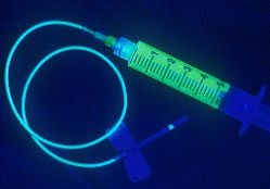
You will be given eye drops that make your pupil dilate. You will be asked to place your chin on a chin rest and your forehead against a support bar to keep your head still during the test. The nurse or photographer will take pictures of the inside of your eye. After the first group of pictures is taken, a dye called fluorescein is injected into a vein. Most often it is injected at the inside of your elbow. A camera-like device takes pictures as the dye moves through the blood vessels in the back of your eye.
How to Prepare for the Test
You will need someone to drive you home. Your vision may be blurry for up to 12 hours after the test. You may be told to stop taking medicines that could affect the test results. Tell your health care provide about any allergies, particularly reactions to iodine.
You may be asked to sign an informed consent form. You must remove contact lenses before the test. Tell the provider if you may be pregnant.
How the Test will Feel
When the needle is inserted, some people feel slight pain. Other others feel only a prick or sting. Afterward, there may be some throbbing. When the dye is injected, you may have mild nausea and a warm feeling in your body. These symptoms go away quickly most of the time. The dye will cause your urine to be darker. It may be orange in color for a day or two after the test.
Why the Test is Performed
This test is done to see if there is proper blood flow in the blood vessels in the two layers in the back of your eye (the retina and choroid). It can also be used to diagnose problems in the eye or to determine how well certain eye treatments are working.
Normal Results
A normal result means the vessels appear a normal size, there are no new abnormal vessels, and there are no blockages or leakages.
What Abnormal Results Mean
If blockage or leakage is present, the pictures will map the location for possible treatment. An abnormal value on a fluorescein or ICG angiography may be due to:
•Blood flow (circulatory) problems, such as blockage of the arteries or veins
•Cancer
•Diabetic or other retinopathy
•High blood pressure
•Inflammation or edema
•Macular degeneration
•Microaneurysms - enlargement of capillaries in the retina
•Tumors
•Swelling of the optic disc
•Retinal detachment
•Retinitis pigmentosa
Risks
There is a slight chance of infection any time the skin is broken. If your arm becomes red, swollen or tender after the test, notify us immediately. The dye may leak outside of the vein and cause discomfort. This is treated with ice pack and Tylenol and will dissipate within a day or two. Rarely, a person is overly sensitive to the dye and may experience:
•Dizziness or faintness
•Dry mouth or increased salivation
•Hives
•Increased heart rate
•Metallic taste in mouth
•Nausea and vomiting
•Sneezing
•Difficulty breathing or wheezing
Serious allergic reactions are rare occurring in about 1/250,000.

Indocyanine Green Angiography (ICG)
The Retina Group of New York utilizes state-of-the-art diagnostic laser scanning ophthalmoscopes to image the retina and choroidal circulation. Following an intravenous injection of a fraction of a teaspoon of a green dye in your arm vein, a low power invisible laser is used to scan the retina and capture high resolution digital images of the retina lining the back wall of the eye. As the dye fills the blood vessels of the two circulatory systems of the eye, the images immediately appear showing abnormal blood vessels or patterns characteristic of many ocular conditions such as macular degeneration, central serous retinopathy and macroaneurysms. The low powered infrared laser can often see through cataracts and blood that fluorescein angiography cannot visualize and confirm suspected pathology. All images are displayed on large monitors and explained so that patients can readily see the progress of their treatments.

